Hip Dysplasia
All NyaStar Irish Red and White Setters are Tested by OFA Good or Excellent.
Click here to see an international comparison Chart
Hip dysplasia literally means an abnormality in the development of the hip joint. It is characterized by a shallow acetabulum (the "cup" of the hip joint) and changes in the shape of the femoral head (the "ball" of the hip joint). These changes may occur due to excessive laxity in the hip joint. Hip dysplasia can exist with or without clinical signs. When dogs exhibit clinical signs of this problem they usually are lame on one or both rear limbs. Severe arthritis can develop as a result of the malformation of the hip joint and this results in pain as the disease progresses. Many young dogs exhibit pain during or shortly after the growth period, often before arthritic changes appear to be present. It is not unusual for this pain to appear to disappear for several years and then to return when arthritic changes become obvious.
Dogs with hip dysplasia appear to be born with normal hips and then to develop the disease later. This has led to a lot of speculation as to the contributing factors which may be involved with this disease. This is an inherited condition, but not all dogs with the genetic tendency will develop clinical signs and the degree of hip dysplasia which develops does not always seem to correlate well with expectations based on the parent's condition. Multiple genetic factors are involved and environmental factors also play a role in determining the degree of hip dysplasia. Dogs with no genetic predisposition do not develop hip dysplasia.
At present, the strongest link to contributing factors other than genetic predisposition appears to be to rapid growth and weight gain. In a recent study done in Labrador retrievers a significant reduction in the development of clinical hip dysplasia occurred in a group of puppies fed 25% less than a control group which was allowed to eat free choice. It is likely that the laxity in the hip joints is aggravated by the rapid weight gain.
If feeding practices are altered to reduce hip dysplasia in a litter of puppies, it is probably best to use a puppy food and feed smaller quantities than to switch to an adult dog food. The calcium/phosphorous to calorie ratios in adult dog food are such that the puppy will usually end up with higher than desired total calcium or phosphorous intake by eating an adult food. This occurs because more of these foods are necessary to meet the caloric needs of puppies, even when feeding to keep the puppy thin.
If clinical signs of hip dysplasia occur in young dogs, such as lameness, difficulty standing or walking after getting up, decreased activity or a bunny-hop gait, it is often possible to help them medically or surgically. X-ray confirmation of the presence of hip dysplasia prior to treatment is necessary. There are two techniques currently used to detect hip dysplasia, the standard view used in Orthopedic Foundation for Animals (OFA) testing and X-rays (radiographs) utilizing a device to exaggerate joint laxity developed by the University of Pennsylvania Hip Improvement Program (PennHIP). The Penn Hip radiographs appear to be a better method for judging hip dysplasia early in puppies, with one study showing good predictability for hip dysplasia in puppies exhibiting joint laxity at 4 months of age, based on PennHIP radiographs.
Once a determination is made that hip dysplasia is present, a treatment plan is necessary. For dogs that exhibit clinical signs at less than a year of age, aggressive treatment may help alleviate later suffering. In the past a surgery known as a pectineal myotomy was advocated but more recent evidence suggests that it is an ineffective surgical procedure. However, administration of glycosaminoglycans (Adequan Rx) may help to decrease the severity of arthritis that develops later in life. Surgical reconstruction of the hip joint (triple pelvic osteotomy) is helpful if done during the growth stages. For puppies with clinical signs at a young age, this surgery should be strongly considered. It has a high success rate when done at the proper time.
Dogs that exhibit clinical signs after the growth phase require a different approach to treatment. It is necessary to determine if the disorder can be managed by medical treatment enough to keep the dog comfortable. If so, aspirin is probably the best choice for initial medical treatment. Aspirin/codeine combinations, phenylbutazone, glycosaminoglycosans and corticosteroids may be more beneficial or necessary for some dogs. It is important to use appropriate dosages and to monitor the progress of any dog on non-steroidal or steroidal anti-inflammatory medications due to the increased risk of side effects to these medications in dogs. If medical treatment is insufficient then surgical repair is possible.
The best surgical treatment for hip dypslasia is total hip replacement. By removing the damaged acetabulum and femoral head and replacing them with artificial joint components, pain is nearly eliminated. This procedure is expensive but it is very effective and should be the first choice for treatment of severe hip dyplasia whenever possible. In some cases, this surgery may be beyond a pet owner's financial resources. An alternative surgery is femoral head ostectomy. In this procedure, the femoral head (ball part of the hip joint) is simply removed. This eliminates most of the bone to bone contact and can reduce the pain substantially. Not all dogs do well following FHO surgery and it should be considered a clear "second choice".
Hip dysplasia may not ever be eliminated by programs designed to detect it early unless some effort is made to publish the results of diagnostic tests such as the OFA evaluation or PennHIP evaluations, openly. This is the only way that breeders will be able to tell for certain what the problems have been with hip dysplasia in a dog's ancestry.
When an older dog is exhibiting signs of pain associated with this condition it is often possible to help them dramatically through medication and simple steps like providing a warm bed or warm spot to rest during the day. There is no advantage to pain and steps should be taken to ensure that the older dog is not in pain. Regular exercise can be very helpful and weight loss can have dramatic effects on the amount of discomfort a dog experiences.
Working with your vet to come to the best solution for your dog and your situation will enable you and your dog to enjoy life to its fullest, despite the presence of hip dysplasia.
OFA TESTING PROCESS
When a radiograph arrives at the OFA, the information on the radiograph is checked against information on the application. The age of the dog is calculated, and the submitted fee is recorded. The board-certified veterinary radiologist on staff at the OFA screens the radiographs for diagnostic quality. If it is not suitable for diagnostic quality (poor positioning, too light, too dark or image blurring from motion), it is returned to the referring veterinarian with a written request that it be repeated. An application number is assigned.
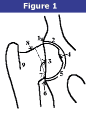
 Radiographs of animals 24 months of age or older are independently evaluated by three randomly selected, board-certified veterinary radiologists from a pool of 20 to 25 consulting radiologists throughout the USA in private practice and academia. Each radiologist evaluates the animal's hip status considering the breed, sex, and age. There are approximately 9 different anatomic areas of the hip that are evaluated (Figure 1).
- Craniolateral acetabular rim
- Cranial acetabular margin
- Femoral head (hip ball)
- Fovea capitus (normal flattened area on hip ball)
- Acetabular notch
- Caudal acetabular rim
- Dorsal acetabular margin
- Junction of femoral head and neck
- Trochanteric fossa
The radiologist is concerned with deviations in these structures from the breed normal. Congruency and confluence of the hip joint (degree of fit) are also considered which dictate the conformation differences within normal when there is an absence of radiographic findings consistent with HD. The radiologist will grade the hips with one of seven different physical (phenotypic) hip conformations: normal which includes excellent, good, or fair classifications, borderline or dysplastic which includes mild, moderate, or severe classifications.
Seven classifications are needed in order to establish heritability information (indexes) for a given breed of dog. Definition of these phenotypic classifications are as follows:
- Excellent
- Good
- Fair
- Borderline
- Mild
- Moderate
- Severe
The hip grades of excellent, good and fair are within normal limits and are given OFA numbers. This information is accepted by AKC on dogs with permanent identification and is in the public domain. Radiographs of borderline, mild, moderate and severely dysplastic hip grades are reviewed by the OFA radiologist and a radiographic report is generated documenting the abnormal radiographic findings. Unless the owner has chosen the open database, dysplastic hip grades are closed to public information.
Hip Grading System with OFA
The phenotypic evaluation of hips done by the Orthopedic Foundation for Animals falls into seven different categories. Those categories are normal (Excellent, Good, Fair), Borderline, and dysplastic (Mild, Moderate, Severe). Once each of the radiologists classifies the hip into one of the 7 phenotypes above, the final hip grade is decided by a consensus of the 3 independent outside evaluations. Examples would be:
- Two radiologists reported excellent, one good—the final grade would be excellent
- One radiologist reported excellent, one good, one fair—the final grade would be good
- One radiologist reported fair, two radiologists reported mild—the final grade would be mild
The hip grades of excellent, good and fair are within normal limits and are given OFA numbers. This information is accepted by AKC on dogs with permanent identification (tattoo, microchip) and is in the public domain. Radiographs of borderline, mild, moderate and severely dysplastic hip grades are reviewed by the OFA radiologist and a radiographic report is generated documenting the abnormal radiographic findings. Unless the owner has chosen the open database, dysplastic hip grades are not in the public domain.
Excellent
Excellent (Figure 1): this classification is assigned for superior conformation in comparison to other animals of the same age and breed. There is a deep seated ball (femoral head) which fits tightly into a well-formed socket (acetabulum) with minimal joint space. There is almost complete coverage of the socket over the ball.

Good
Good (Figure 2): slightly less than superior but a well-formed congruent hip joint is visualized. The ball fits well into the socket and good coverage is present.

Fair
Fair (Figure 3): Assigned where minor irregularities in the hip joint exist. The hip joint is wider than a good hip phenotype. This is due to the ball slightly slipping out of the socket causing a minor degree of joint incongruency. There may also be slight inward deviation of the weight-bearing surface of the socket (dorsal acetabular rim) causing the socket to appear slightly shallow (Figure 4). This can be a normal finding in some breeds however, such as the Chinese Shar Pei, Chow Chow, and Poodle.


Borderline
Borderline: there is no clear cut consensus between the radiologists to place the hip into a given category of normal or dysplastic. There is usually more incongruency present than what occurs in the minor amount found in a fair but there are no arthritic changes present that definitively diagnose the hip joint being dysplastic. There also may be a bony projection present on any of the areas of the hip anatomy illustrated above that can not accurately be assessed as being an abnormal arthritic change or as a normal anatomic variant for that individual dog. To increase the accuracy of a correct diagnosis, it is recommended to repeat the radiographs at a later date (usually 6 months). This allows the radiologist to compare the initial film with the most recent film over a given time period and assess for progressive arthritic changes that would be expected if the dog was truly dysplastic. Most dogs with this grade (over 50%) show no change in hip conformation over time and receive a normal hip rating; usually a fair hip phenotype.
Mild
Mild Canine Hip Dysplasia (Figure 5): there is significant subluxation present where the ball is partially out of the socket causing an incongruent increased joint space. The socket is usually shallow only partially covering the ball. There are usually no arthritic changes present with this classification and if the dog is young (24 to 30 months of age), there is an option to resubmit an radiograph when the dog is older so it can be reevaluated a second time. Most dogs will remain dysplastic showing progression of the disease with early arthritic changes. Since HD is a chronic, progressive disease, the older the dog, the more accurate the diagnosis of HD (or lack of HD).

Moderate
Moderate Canine Hip Dysplasia: there is significant subluxation present where the ball is barely seated into a shallow socket causing joint incongruency. There are secondary arthritic bone changes usually along the femoral neck and head (termed remodeling), acetabular rim changes (termed osteophytes or bone spurs) and various degrees of trabecular bone pattern changes called sclerosis. Once arthritis is reported, there is only continued progression of arthritis over time.
Severe
Severe HD (Figure 6): assigned where radiographic evidence of marked dysplasia exists. There is significant subluxation present where the ball is partly or completely out of a shallow socket. Like moderate HD, there are also large amounts of secondary arthritic bone changes along the femoral neck and head, acetabular rim changes and large amounts of abnormal bone pattern changes.

